Imaging of the body can help doctors understand what is happening inside and plan what treatment is needed. The Imaging Department offers a range of diagnostic imaging and interventional radiology services for children and young people up to 16 years of age.
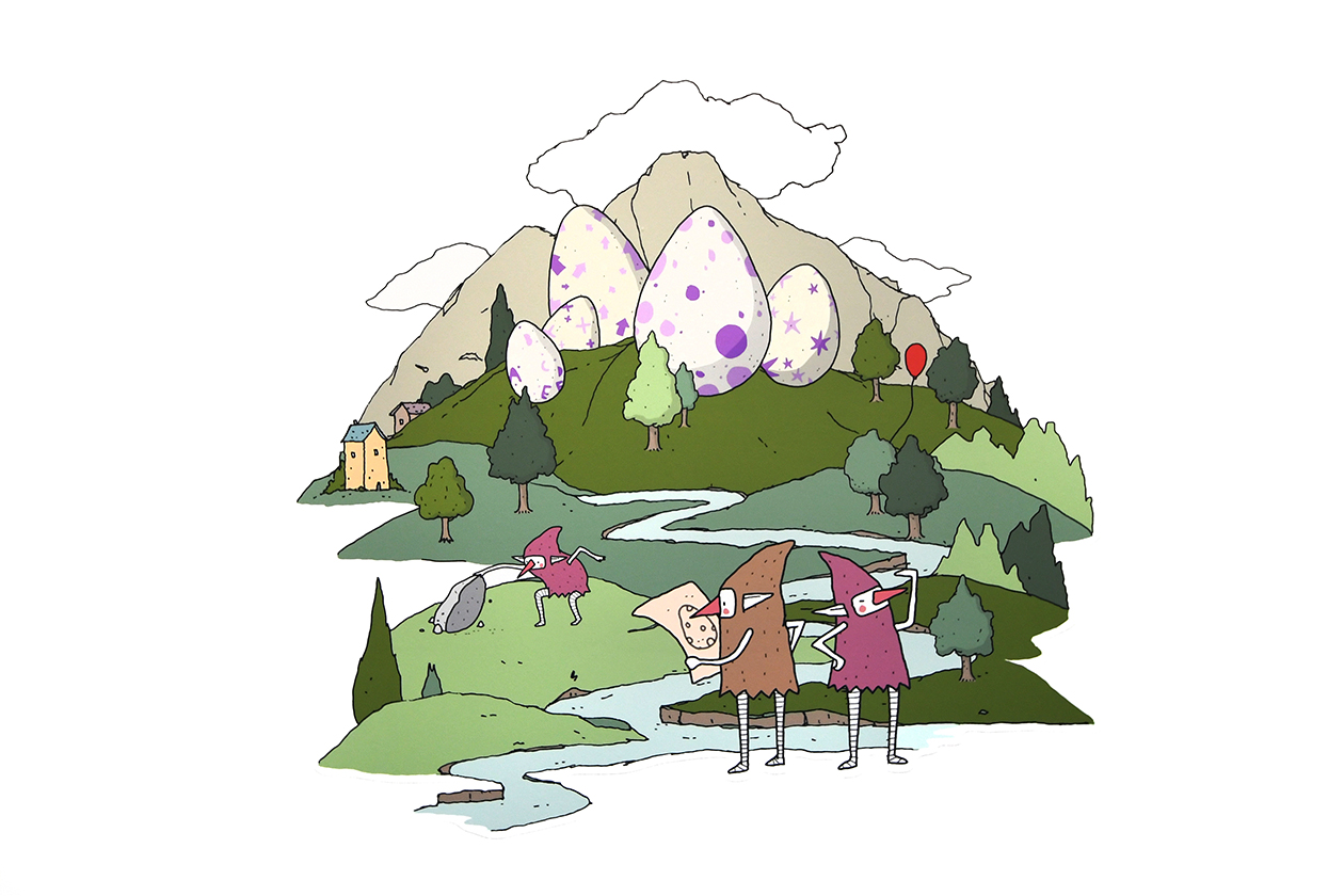
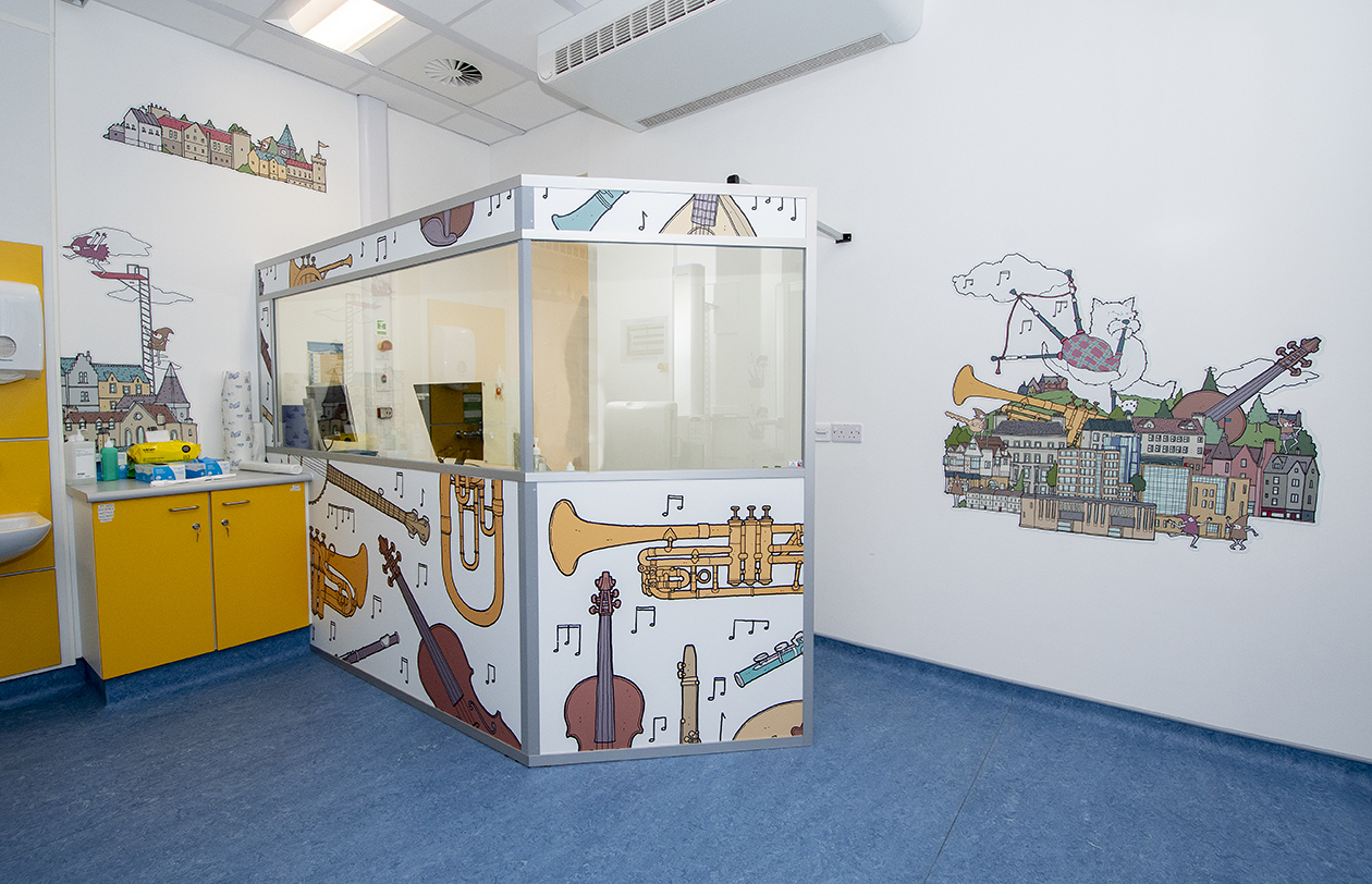
If you have an appointment for a scan or x-ray and have been asked to come to the Imaging Department, it can be found on the ground floor, on the right-hand side, after entering through the main RHCYP entrance. There is a reception desk for booking in on arrival. Sometimes families have a short wait, so our waiting area has toys and other play equipment for children to explore and enjoy. See if you can spot our cheeky brownies in the artwork all around the walls.

Tests, scans and x-rays
There are different tests, scans and x-rays that can show different views and functions of the body. The type you need will be ordered by the doctor or nurse in charge of each young patient’s care. The tests we perform include general x-rays, fluoroscopy (x-ray screening), ultrasound, nuclear medicine, computerised tomography (CT), magnetic resonance imaging (MRI). The reason for the type of test needed will be explained to patients and their parents and carers. Our staff are always happy to answer questions and help children relax as much as possible during a test, scan or x-ray.
X-ray Internal page link
X-ray machines use radiation to generate pictures of inside the body – most commonly used to look at bones; they can also show other parts of the body, like the chest & lungs. At the RHCYP, there are two digital x-ray rooms, three mobile Image Intensifiers, four mobile x-ray machines and a dental x-ray unit (for teeth). The Emergency Department also has its own x-ray room so that patients can have quick access to an x-ray if it is needed.
Fluoroscopy Internal page link
Fluoroscopy uses low-dose x-rays to create “real-time” moving images of inside the body. It helps doctors see how the body functions. The Fluoroscopy room is equipped with a Seimens Artis Zee fluoroscopy suite.

Ultrasound Internal page link
An ultrasound scan uses sound waves to show pictures of the inside of the body. There are two ultrasound rooms equipped with GE Logiq ultrasound machines.

CT Scan Internal page link
Computerised Tomography (CT scans) create 3D images of the body using x-rays. A CT scan can show different layers, such as bones, organs, blood is moving around the body, and give a complete picture of injuries or illnesses. The Department has a Canon Aquilion One, 64 slice CT scanner.

Nuclear Medicine Internal page link
A nuclear medicine scan involves a small injection of a special radioactive “dye”, showing up on a gamma camera. It can show how well different organs in the body are working. Nuclear medicine has a GE SPECT Gamma Camera (Discovery 670).
MRI Internal page link
The initials MRI stand for magnetic resonance imaging. This type of scan uses Magnetic fields, radio waves and computers to show a 3D picture of almost any part of the body. It does not use any x-rays. The MRI department has two Philips Ingenia 3.0T scanners, including an intra-operative scanner – this is a scanner located next to the operating theatre so that patients can have an MRI scan immediately after their operation. The Philips Patient Ambient Lighting system and kitten scanner are interactive tools that can make the examination more fun and less stressful for children.
X-Ray
What is an x-ray?
An x-ray is the word we use to describe a picture taken by an x-ray machine. X-ray radiation and a detector are used to make an image of the inside of your child’s body.
What do x-ray images look like?
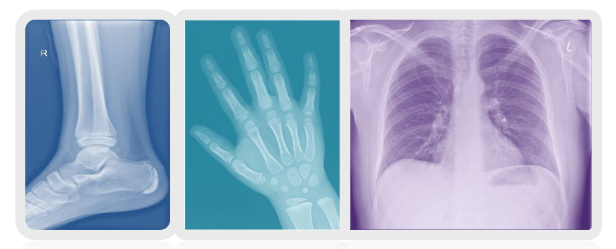
X-ray images are black and white pictures with lots of shades of grey in between. The shades of grey depend on the density of the object being x-rayed. In the human body, bones are more dense than muscles which are in turn, more dense than the air in your lungs. The dense parts of your body look lighter and the less dense parts look darker.
 |
 |
|---|
Why are x-rays used?
X-rays help the doctor or health care practitioner either make a diagnosis or monitor the progress of your child’s treatment. We ensure that when anyone has an x-ray, the benefits from making the right diagnosis or providing the correct treatment outweigh the very low risk of having the x-ray itself.
What will it involve?
You will be taken to the x-ray room where you will meet the radiographer. Depending on which part of their body is to be x-rayed, your child may need to be undressed. The department will provide a gown for older children to wear. It is important that your child is still for the x-ray. You can stay with your child to comfort and distract them and when necessary help to hold them in the correct position.
Pregnancy:
There are safety rules that we follow regarding pregnant women. If you are or may be pregnant, please tell the radiographer.
Can you feel an x-ray?
You can not feel, see or hear an x-ray. An x-ray does not hurt.
Information about x-ray radiation:
We are all exposed to natural background radiation every day of our lives. This comes from our environment, the air we breathe, the food we eat (bananas and brazil nuts for example) and even from outer space (cosmic rays). In Scotland, natural radiation comes from underlying rocks and building materials such as granite. Over a year, our exposure will vary according to where we have lived, where we have flown to and what we have eaten.
Each medical x-ray gives a small additional dose on top of this natural background radiation. The level of dose varies with the type of procedure. Common x-rays such as the chest or limbs (arms and legs) involve small amounts of radiation that are similar to less than 3 weeks of natural background radiation.

Xrays explained for Children

Fluoroscopy
Why does your child need a fluoroscopy scan?
While other scans can show the size and shape of part of your child’s body, a fluoroscopy scan helps provide information about the structure, location or function of different parts of the body. This can help with diagnosis and treatment planning.
How does fluoroscopy work?
Fluoroscopy uses low-dose x-rays to create “real-time” moving images of inside the body. It helps doctors see how the body functions. Often a liquid contrast will be given to highlight regions of interest. The exact method for your child’s fluoroscopy will vary depending on their specific examination.

What will the test involve?
You will be taken to the x-ray room, where you will meet the radiographer and radiologist. They will explain the test, and there will be an opportunity to ask any questions. If possible, children should wear a plain top, as some designs or motifs will show up on the pictures. If your child needs to get changed then the department will provide a gown.
X-ray pictures will be taken with your child in different positions but this will be explained clearly to them at the time. You will be asked to stay in the fluoroscopy room with your child.
Fluroscopy & pregnancy
If you are or could be pregnant, you will not be able to stay in the room while the scan is being done. Another parent or carer should stay with your child in these circumstances. Other children will not be allowed in the scanning room so please make arrangements for them to be looked after at home.
What if my child is taking medication?
Do not stop taking any prescribed medicines – the scan will not make them less effective. If you have any questions about this please contact the department before you come, or ask on the day.
Are there any risks involved?
Fluoroscopy scanners use radiation; x-rays generate the pictures we need for each child’s diagnosis and treatment. The amount of radiation is kept as low as possible to get the pictures needed.
We are all exposed to background radiation every day. Medical radiation gives us an additional dose which varies with each examination. Your child’s doctor has asked for this scan as they feel that the benefits of the procedure outweigh any risk of having the scan.
Fluoroscopy explained for Children

Ultrasound
What is an Ultrasound Scan?
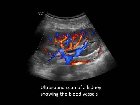
An ultrasound scan uses sound waves to show inside the body. This type of test does not use x-rays.
 |
 |
 |
|---|
What will the test involve?
When your child arrives they will be taken to the ultrasound room where they will be met by a department assistant and the sonographer or doctor. The ultrasound scan will be explained and there will be plenty of time to ask questions. You are able to stay with your child whilst this test is being carried out.
Your child will be asked to lie on the ultrasound couch. Some warm jelly will be put on the area that is being scanned. This is used to give the doctor a better picture and can be a little sticky but will be wiped off at the end. The doctor will use a small hand probe and move it gently over the area looking at the pictures on a screen.
Will the test hurt?
This scan should not hurt. If the area being scanned is tender then there may be a little discomfort but the doctor will be as gentle as they can.
How long will it take?
This test will take 10-15 minutes.
Does the scan require specific preparation?
It would be useful if your child could be dressed in loose clothing that can be moved easily from the area that is to be scanned.
Some scans may need your child to fast for a period of time before the scan or we may ask for your child to attend with a full bladder – this will be included in your child’s appointment letter if it applies to them.
Information about fasting
The most common reason for not feeding before an ultrasound scan is to help us get pictures of the gallbladder and the related ducts in the liver.
If your child is diabetic the period of fasting may need to be adjusted. Please contact the department.
If your child is taking medication, please give it to them as usual but avoid giving milk when doing so.
If you are uncertain what to do please contact the department.
If you think your child can cope with a period of fasting but the period suggested in the instructions is just a little bit longer than you think they will manage then please use your judgement – we are aiming for the longest period of fasting achievable which will not cause your child distress.
Information on bladder filling
In your child’s appointment letter, we may have asked for your child to attend with a full bladder.
If your child has not yet begun or is still in the process of toilet training you can disregard this information.
We appreciate that younger children, even if fully toilet trained, can find it difficult to get the timing right. We are also aware that older children may have an issue with the bladder that make it difficult to fill on-demand.
If you think it might be difficult for your child to fill their bladder for your appointment time (bearing in mind that appointment times sometimes run a little late), our advice would be to not make a big effort to fill the bladder up. We can do a quick scan to see if the bladder is full enough to scan. If not, you can wait nearby with a drink of water and can usually come back to the ultrasound room without a long delay.
What if my child is taking medication?
Do not stop taking any prescribed medications, as it will not affect your scan. The scan will not affect how well any medicines work. If you have any questions about this do not hesitate to contact the department.
Ultrasound explained for children

CT Scan
Why does your child need a CT scan?
While different scans can show the size and shape of a part of your child’s body, CT gives a much more detailed picture. This can help with diagnosis and treatment planning.
What is a CT scanner and how does it work?
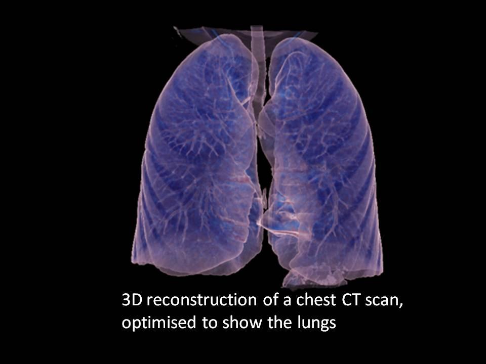
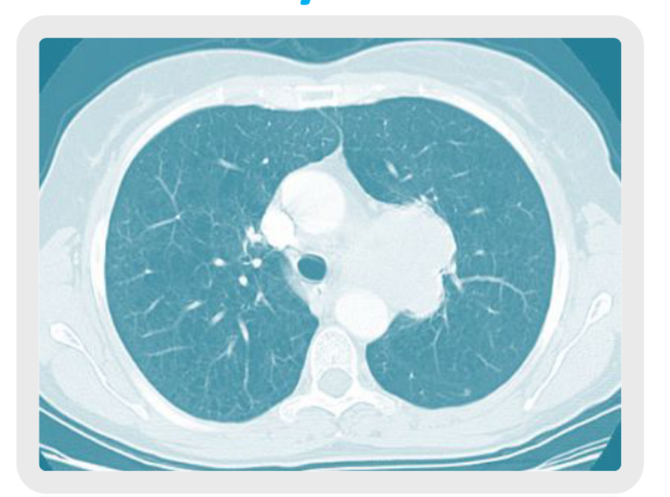
The initials CT stand for Computerised Tomography. CT is a method of obtaining high-quality 3D images using x-rays. Multiple images or “slices” are taken at different angles which are then processed by a computer to produce a 3D picture of the body. It can image both bone and soft tissue.
 |
 |
|---|
What will the CT scan involve?
Your child will be positioned on a bed that moves into the scanner as it takes the scan. The CT scanner looks rather like a large polo-mint and sounds a bit like a washing machine. It is important that your child keeps very still.
The radiographer will be able to see your child and talk to them through a two-way intercom at all times throughout the scan which typically takes 5 – 10 minutes. You will be asked to stay in the scanning room with your child.
Pregnancy & CT scans
If you are or may be pregnant you will not be able to stay in the room while the scan is being done. In this case, another parent or carer should stay with your child. Other children will not be allowed in the scanning room so please make arrangements for them to be looked after at home.
Will my child need an injection?
Your child may need to have an injection of contrast (x-ray dye) which shows up in the organs and blood vessels. The needle and tube through which we give the contrast are called cannula. We can use skin numbing cream to hopefully help with any discomfort when the needle is put in. This is sometimes known as ‘magic cream’.
If an injection of dye is needed, we will ask about any medical history of allergies, diabetes, asthma or kidney problems. The dye may leave a metallic taste in the mouth and a warm sensation through the body.
What if my child is taking medication?
Do not stop any prescribed medications. The scan will not affect how well they work. If you have any questions about this please contact the department.
Are there any risks involved?
CT scanners use radiation – x-rays to generate the 3D pictures we need for your diagnosis and treatment; the amount of radiation we use is kept as low as possible to get the pictures we need.
We are all exposed to background radiation every day. Medical radiation gives us an additional dose which varies with each examination. Your child’s doctor has asked for this scan as they feel that the benefits of the procedure outweigh any risk of having the scan.
What happens afterwards?
Once the examination is complete, your child will be able to leave the department. If they have had an injection of contrast dye they will be asked to stay in the department for a further 15 minutes. Your child should not experience any discomfort following their CT scan and can return to their normal daily routine.
Nuclear Medicine
Why does your child need a nuclear medicine scan?
While other scans can show the size and shape of a part of your child’s body, a nuclear medicine scan helps provide information about the structure, location or function of different parts of the body. This can help with diagnosis and treatment planning.
What is a nuclear medicine scan and how does it work?
A special radioactive liquid called an isotope is injected into a vein. This liquid will travel to the specific area of the body we are examining, it will emit gamma rays that are recorded by the gamma camera and analysed by a computer to create a picture. This allows us to see what’s inside the body.

What will the test involve?
When you arrive you will meet the radiographer and radiography assistant. They will explain the test, and there will be an opportunity to ask questions. Your child will be weighed and then will be given a small injection of radioactive liquid. If your child has a favourite toy, they can bring it along with them.
Pictures are taken using the gamma camera. There sometimes needs to be a delay between the injection and the pictures being taken. Depending on the type of scan, the imaging can be immediate or there might be a delay of between 90 minutes to 24 hours). In cases like this, the radiographer will give you time to come back to the department for the pictures.
Different sets of pictures are taken, with your child lying still on the bed. Your child will not need to undress. They can watch a DVD, listen to music, or we have distraction toys for younger children. You can stay with your child during the test and help to keep them still.
Will the test hurt?
Your child will not feel any discomfort from the radioactive liquid or imaging process. An injection is needed for this examination. The needle used for the injection is very small and will only be in for a very short time.
Are there any risks involved?
A nuclear medicine scan involves an injection of a radioactive isotope – the amount of radiation we use is kept as low as possible to get the pictures we need. We are all exposed to background radiation every day. Medical radiation gives us an additional dose which varies with each examination. Your child’s doctor has asked for this scan as they feel that the benefits of the procedure outweigh any risk of having the scan.
Precautions if you are pregnant or breastfeeding.
If you are pregnant, it would be best if someone else could accompany you and your child for the test. We would recommend avoiding close contact (prolonged cuddles) for 24 hours after the injection has been given. Breastfeeding can continue as normal.
What if my child is taking medication?
Do not stop taking any prescribed medications. The scan will not affect how well they work.
What happens afterwards?
Once the test is over, you will be able to leave the department.
Your child should avoid sharing a bath with other children on the night of the test. Any wet nappies should be disposed of in an outside bin.
Your child can return to school or nursery the following day.
Nuclear Medicine Test
Nuclear Medicine explained for children

MRI
Why does your child need an MRI scan?
While different scans can show the size and shape of a part of your child’s body, MRI gives a much more detailed picture. CT & X-rays are good for showing the outline of bone. MRI & ultrasound are good for the soft tissue within your skeleton. This can help with diagnosis and treatment planning.
What is an MRI scanner and how does it work?
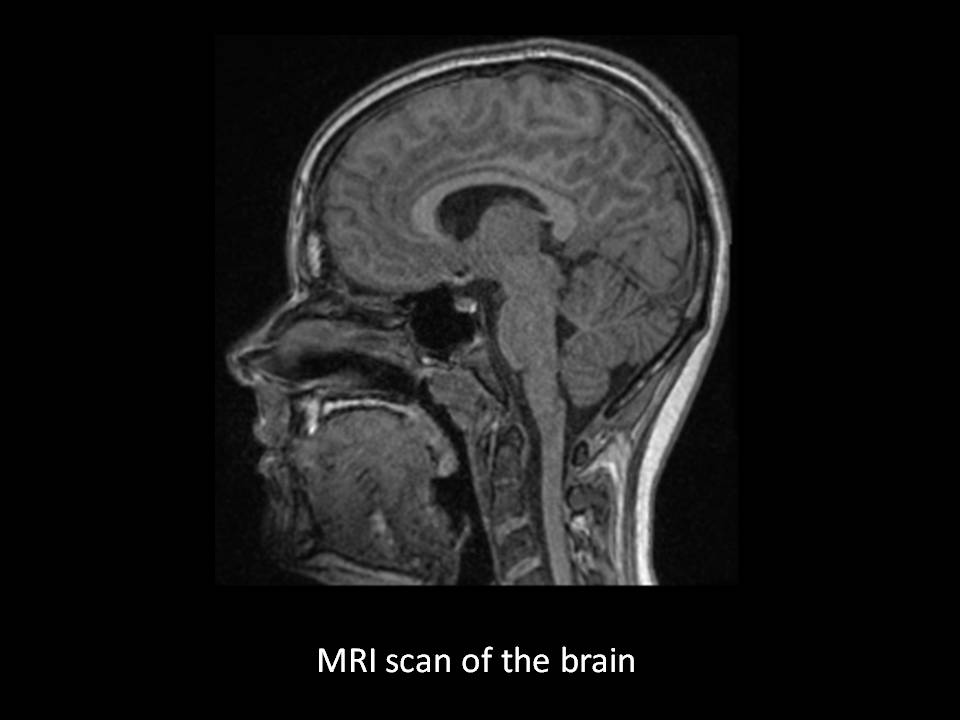
The initials MRI stand for Magnetic Resonance Imaging. Magnetic fields, radio waves and computers are used to produce picture ‘slices’ of the human body. It does not use any x-rays and is considered completely safe.
 |
 |
|---|
What will the MRI examination involve?
Your child will lie on a bed within the scanner and will be closely monitored by the radiographer. They will feel no effects during this scan. The scanner makes a knocking sound (sometimes like a digger noise) so ear protection will be placed on your child. You can bring a DVD with you for your child to listen to or watch during the scan (we also have a large selection of films). The radiographer will be able to see your child at all times during the scan, which can take between 15-60 minutes. Sometimes your child will need an injection of a contrast agent, to help the doctor see parts of the body more clearly and aid diagnosis. The MRI bed moves a little – or a lot – to get your child in the right place. Sometimes this happens with little or no warning.
You will be able to stay in the scanning room with your child. If you are or may be pregnant or have any yes responses listed under important safety information on the safety checklist, you will not be able to stay in the room while the scan is being done. In this case, another parent or carer can stay with your child. Other children will not be allowed in the scanning room so please make arrangements for them to be looked after at home.
An exciting addition to our MRI department at RHCYP is a kitten or ‘mini’ MRI scanner. This is an interactive play therapy tool in which your child can scan toys, learn how the scanner works and see the toys ‘insides’ making the experience fun and less scary. The themes seen in the kitten scanner can be carried through into the scan room with the use of our new ambient lightning experience. Your child can choose a theme or colour for their scan, creating a less stressful and more fun environment. Being able to personalise the experience helps children feel more in control.
Watch Basil Brush meet some of the MRI team at the hospital in Edinburgh and find out all about it!
Having an MRI under General Anaesthetic
Some babies, children or young people will have a general anaesthetic before their MRI scan. This means having some sleep medicine and going to sleep for a short time while the scan takes place.
If you are having this type of procedure, a letter will be sent with specific details with instructions about the time to arrive, as well as the place, date and information about fasting. Download the PDF file of the ‘MRI scan under general anaesthetic’ leaflet.
Find out about having an anaesthetic.
MRI videos
What happens in MRI
Prepare for MRI
Check out Philips – Kitten scanner
Also check out Philips Ingenia scanners – Ambient lighting system
Will my child need an injection?
Your child may need to have an injection of contrast that shows up in the organs and blood vessels. The needle and tube through which we give the contrast are called cannula. We can use skin numbing cream which helps with any discomfort when the needle is put in. This is known as ‘magic cream’.
What if my child is taking medication?
Do not stop any prescribed medications. The scan will not affect how well they work. If you have any questions, please contact the department.
Important safety information
MRI uses a strong magnet. It may not be possible to have an MRI scan if your child has:
- A heart pacemaker
- Certain inner-ear implants
- Metal fragments within their eyes or head
- Surgical clips within their head
- Neuro-electrical stimulators
- some other implants e.g. artificial heart valves, orthopaedic implants or plate. (hip replacements are generally safe).
Please contact the MRI unit if you or your child has any of the above or has a programmable shunt. You will receive a safety questionnaire with your appointment letter, please complete the questionnaire for your child and return it when attending your appointment. For an MRI scan, you Must Remove Items – metal objects such as watches, removable dental plates or braces, hearing aids, mascara, jewellery, including piercings, must be removed. A locker will be provided for personal items, however, it is better if you can leave these at home.
What happens afterwards?
Once the examination is complete, your child will be able to leave the department. Your child should not experience any discomfort following their scan and can return to their normal daily routine.
MRI explained for children

What we do & Our Team
There are 24 radiographers, a sonographer, 7 radiology consultants, 6 radiology department assistants and 3 administration staff members in the Imaging Department team. The RHCYP is also a teaching hospital, so you might also meet radiology trainees and student radiographers who come for specialist training. Everyone will introduce themselves and give their names when they meet patients and their families.
- A radiologist is a medical doctor who specialises in diagnosing and treating disease and injury using imaging techniques such as X-rays, computed tomography (CT), magnetic resonance imaging (MRI), nuclear medicine, and ultrasound.
- A diagnostic radiographer specialises in taking images of the body Opens in new window for the diagnosis Opens in new window and treatment Opens in a new window of pathology Opens in a new window. They are trained in a range of imaging techniques, including X-ray, Ultrasound, Fluoroscopy, Computerised Tomography (CT), Magnetic Resonance Imaging (MRI), and Nuclear Medicine.
- A sonographer is a radiographer who specialises in ultrasound scanning.
- A radiology department assistant (RDA) works closely with all radiology team members, helping them and looking after the patient and family while tests are being carried out.
Meet the team
Medical Imaging is a busy department, with many staff working shifts so that babies, children and young people can have the scans and x-rays that they need, including in an emergency. Key personnel in our department include:
Lead Clinician
Dr Alan Quigley
Consultant Radiologists
Dr Jeremy Jones
Dr Tom Foster
Dr Rachael Kirkbride
Dr Alan Quigley
Dr Michael Jackson
Dr Andrew Kirby
Dr Samantha Choi
Assistant Directorate Manager
Carol Stewart
Principal Radiographer
Louisa Armstrong
Radiographer Team Lead
Jayne Davison
Deputy Team Lead/ CT Lead
Claire Donoghue
Alison Roger
Administrator Lead
Nicola Fleming
